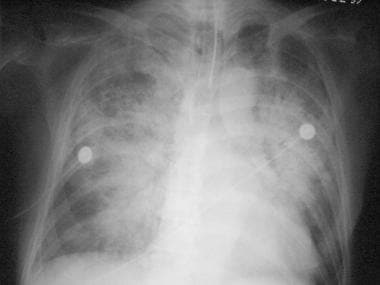
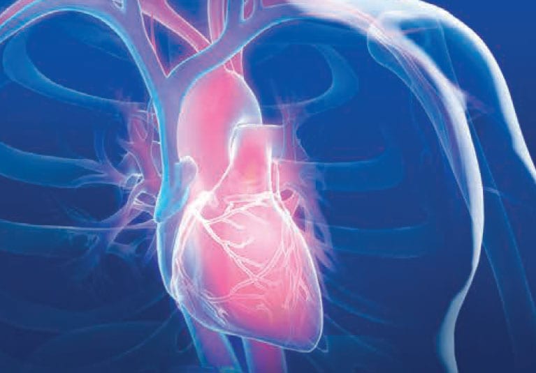
And over time a series of cardiac images can help track the results of treatment or the progression of disease. An advantage of choosing magnetic resonance for cardiac imaging is the free choice in obtaining imaging planes of cardiovascular anatomy in any arbitrary view since this technique is not hampered by the limited availability of acoustic windows as with ultrasound.
 The Science Behind X Ray Imaging
Nicer series on diagnostic imaging.

(PDF) Chest And Cardiac Radiology Nicer Series On Diagnostic Imaging Download. Nicer series on diagnostic imaging. The successful interpre tation of thoracic imaging studies requires the recognition and understanding of the radiologic signs that are charac teristic of many complex disease processes. A cardiac radiologist supervises or performs and then interprets medical images to diagnose diseases of the heart such as heart disease leaky heart valves and defects in the size and shape of the heart.
Usually of an arm or a leg and then threaded through the circulatory system to the heart. Chest and cardiac radiology. Cardiac imaging is a subspecialty of diagnostic radiology.
The lateral chest view may be performed as an adjunct to a frontal chest radiograph in cases where there is diagnostic uncertainty. Gastrointestinal and urogenital radiology. Start studying radiology and diagnostic imaging.
With the advent of echocardiography and cardiac ct and mri the role of chest x rays in evaluating congenital heart disease has been largely been relegated to one of historical and academic interest although they continue to crop up in radiology. Gastrointestinal and urogenital radiology. Critical in the complete evaluation of the cardiac and.
Learn vocabulary terms and more with flashcards games and other study tools. Chest and cardiac radiology. Conclusion the educational objectives for this case based self assess.
Nicer series on diagnostic imaging. Nicer series on diagnostic imaging. Cardiac imaging also helps determine damage from a myocardial infarction or heart attack.
A chest x ray is a visualization of the interior of the chest. Nicer series on diagnostic imaging. Collaborating with the patient and.
Chest imaging remains one of the most complicated sub specialties of diagnostic radiology. Nicer series on diagnostic imaging. Nicer series on diagnostic imaging.
Congenital heart disease chest x ray an approach dr henry knipe and aprof frank gaillard et al. Nicer series on diagnostic imaging. The lateral chest view examines the lungs bony thoracic cavity mediastinum and great vesselslateral radiographs can be particularly useful in assessing the retrosternal and retrocardiac airspaces.
The cardiac and pulmonary imaging section at ucsf radiology is dedicated to safely performing the most current clinical imaging exams of both the respiratory and cardiovascular systems using advanced imaging modalities such as detailed cta and ct exams. Nicer series on diagnostic imaging. Many patients who use this subspecialty have unexplained chest pain or chest pain when exercising angina.
 Thorax Radiography An Overview Sciencedirect Topics
 Diagnostic Imaging Preferred Imaging

 Pin By Kevin Ferrier On Radiologic Technology Radiology
 Diagnostic Imaging Uw Veterinary Care
 Cardiac Mrimra Cedars Sinai
 Cardiac Imaging Tests Cardiovascular Disorders Merck
 How To Read Chest X Rays International Emergency Medicine
X Ray Film Reading Made Easy
 Radiology Quiz 13720 Radiopaediaorg
Chest Radiographs Of Cardiac Devices Part 1 Lines Tubes
 Why The 3 Tesla Mri Is The Best Scanner For Diagnostic
 New Protocols Allow For Mri In Selected Patients With
 Artificial Intelligence Could Revolutionize Medical Care
 How Artificial Intelligence Will Change Medical Imaging

